
In issue 77 of ProAgri Zambia, we presented an overview of the signs and lesions of infectious bronchitis Variant 2 (also known as IS/1494/06) in layer hens. This variant was first reported in Zambia in 2017, and Phibro has detected it again in 2022, confirming its circulation in the country. In this article we revise those concepts. As usual, please consult to your veterinary consultant, he/she is the one to diagnose whether the variant is present at your farm.
What is a variant and why is it important to be aware of its presence?
A variant is a new strain of a given virus that, due to changes in very specific points of its genetic material, presents modifications in its structure and function. This results in changes in its virulence, in the type of disease it produces and, depending on the vaccination plan and vaccines used in the broiler flock, it might be able to escape immunity and infect vaccinated birds.
What do you see in layer birds?
In cases of early infection:
If the infection with IB Variant 2 occurs during the pullet phase, there will be a permanent damage in the reproductive tract. In cases when the virus infects the birds in the first or second week of age, birds may develop silent layer syndrome. In this case, birds reach sexual maturity and, most of the times, they look like a normal adult hen, but they do not produce eggs. Although the ovary is functional and ovulation is verified, the oviduct (Figure 1) will develop cysts in its wall (Figure 2), preventing the progression of the yolk along the oviduct, resulting in failure to produce eggs.
In a few cases, when cysts are very large, the abdomen appears distended, and the birds adopt a penguin-like vertical position. If the birds are in cages, the ones with distended abdomens normally choose to stay at the back of the cage. The egg yolks are released inside the abdomen and reabsorbed, although peritonitis (inflammation and infection in the abdomen) can sometimes develop.
It is difficult to detect false layers, as most of the times they do not show any signs suggesting disease. They are normally found when inspecting the flock due to unusual production curves, for example in flocks where there is a delay to reach peak and/or the maximum production is considerably lower than the production target.
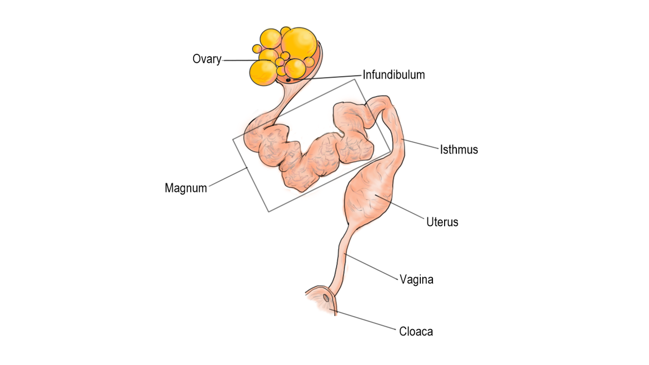
Figure 1: Reproductive tract of the hen (After Jonchere et al., 2010)
When layers get infected:
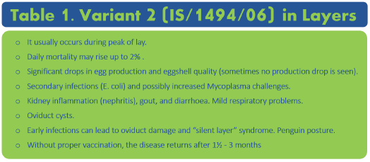
Changes in egg production: Although layers can be infected at any age, infection often occurs during the peak of lay. Infected flocks verify a drop in production sometimes as large as 50%. Usually, egg production recovers in approximately 3 to 4 weeks, however it stabilises at levels lower than before the outbreak. Some flocks do not show production drops. If birds are not vaccinated after the outbreak, using the correct vaccine at the right time, the problem will return every 6 to 12 weeks; this normally results in oscillations of egg production, mostly accompanied by changes in egg quality.
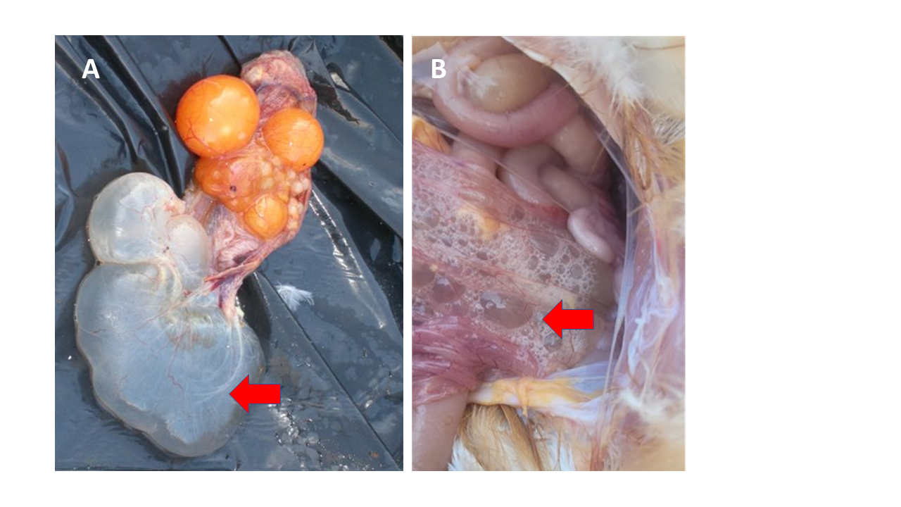
Figure 2: Silent layer syndrome. A and B: red arrow shows cystic oviduct.
Changes in egg quality
The virus affects different sections of the oviduct (Figure 1), resulting in a range of changes in egg quality.
Lesions in the magnum:
The magnum (Figure 1) is the section of the oviduct that secrets the egg albumin. The inflammation of its wall results in eggs with very liquid albumin, or an albumin that liquifies within a couple of minutes after cracking the egg open.
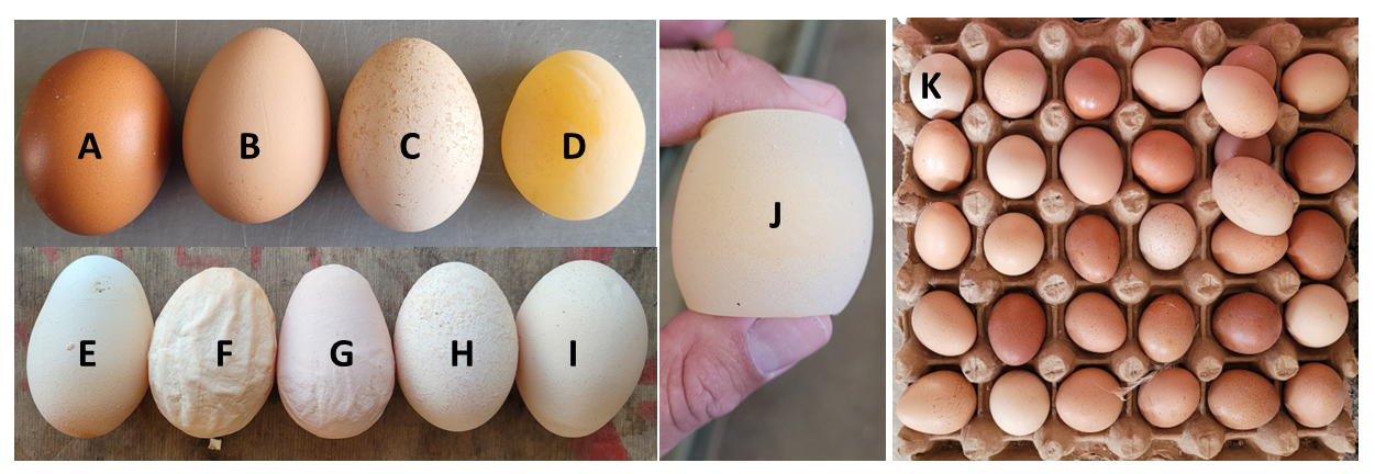
Figure 3: Eggshell alterations due to IB Variant 2. A: Normal; B: Discoloured eggs; C and H: Discoloured shells with calcium deposits; D and J: Soft shell eggs; E, F, and G: Misshapen eggs; I: White, brittle shell; K: Egg tray showing variations in colour and shape.
Lesions of the uterus:
The uterus (Figure 1) is the organ where the shell is formed and the external cuticle, responsible for the egg colour, is deposited.
IB lesions in the uterus initially cause eggs to lose their shine and their traditional golden colour (Figure 3). This translates into a range of different tones of brown when inspecting egg trays after collection (Figure 3). The discolouration may eventually lead to completely white shells (Figure 3).
When the virus affects the uterine glands in charge of producing calcium carbonate, the shells become thinner, brittle, chalky, deformed, or even soft (when the mineralisation is totally absent) (Figure 3). Sometimes, eggs are laid with irregular deposits of calcium on their surface (Figure 3).
As mentioned above, if birds are not vaccinated with the correct vaccine and at the right times, the problems with egg quality and production will return in 6 to 12 weeks.
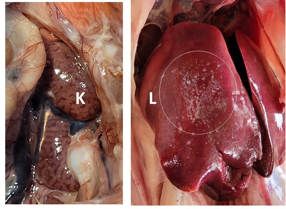
Figure 4: A. Nephritis (kidney inflammation with accumulation of urates) (K: Kidney); B. Urate accumulation on liver surface (gout) (L: Liver).
Other signs and lesions observed
Infected layers can present mild respiratory problems, concurrent with inflammation of kidneys (nephritis), and diarrhoea. When the damage in the kidneys is severe, urates start to accumulate first in the kidneys (Figure 4) and then in other internal organs, condition known as gout (Figure 4). Daily mortality can sometimes be as high as 2%.
The impact on the flock performance can be intensified by the occurrence of secondary infections, especially in flocks infected by mycoplasmas. These birds show generalised infections (septicaemia, polyserositis), airsacculitis (inflammation of airs sacs). This translates into long-haul respiratory symptoms, higher mortality, and low productive performance.
Not everything is IB
There are diseases other than infectious bronchitis producing drop in production and alterations in eggshell quality, such as Newcastle Disease, Egg Drop Syndrome, and Infectious Laryngotracheitis. Furthermore, other IBV strains also can produce qualitative and quantitative issues in production. Therefore, consult your poultry vet as soon as production issues set in.
How to prevent the infection with Variant 2
Phibro TAbic® IB VAR206 is the only vaccine that will give the highest protection against Variant 2, since it contains a virus that is the most similar one to the field strain. The optimum protection is achieved after priming the birds with two doses of the vaccine separated 10 to 14 days from each other, followed by boosters at 10 to 12 weeks, just before transfer, and every 8 weeks in lay (Table 2).
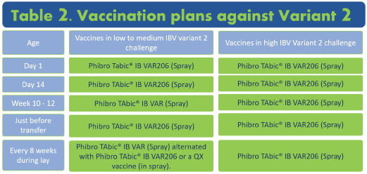
The vaccine SHOULD be applied by coarse spray, or by eye drop; levels of protection achieved with vaccination in drinking water are very poor. The vaccine water should be maintained between 4 and 10 °C, since there is a reduction of a 3 to 3,5 % in effectiveness per degree Celsius of temperature increase above 10 °C.
In order to protect against early Variant 2 infection and avoid the silent layer syndrome, birds SHOULD be spray vaccinated with Phibro TAbic® IB VAR206 at day 1 at the hatchery, followed by a second spray vaccination at the farm at day 12. The vaccine can be sprayed together with Phibro TAbic® V.H. against Newcastle Disease.
As usual, please consult your poultry vet when you suspect your flock is sick. The veterinarian is the only one able to give an appropriate diagnosis and recommend the right treatment and prevention strategies.


